Independently developedhigh-end imaging equipment
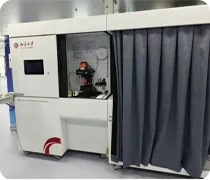
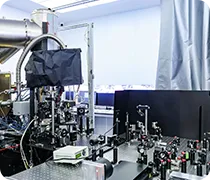
OPM-MEG enables helium-free, non-invasive brain mapping via personalized sensor arrays, delivering millisecond temporal and millimeter spatial precision for brain-function and BCI research.
An integrated five-modality molecular imaging platform for preclinical research and drug development. Combining PET, SPECT, CT, FMT, and MRI in a single system, it enables comprehensive anatomical, functional, and molecular imaging across multiple spatial scales. The platform supports oncology research, drug development, disease mechanism studies, and translational medicine through efficient multimodal in vivo imaging.
The world’s first Total-body TOF PET/CT featuring integrated Depth-of-Interaction (DOI) technology. Featuring a 2-meter axial field of view, the system enables single-bed dynamic whole-body molecular imaging with ultra-high sensitivity, high spatial resolution, and low-dose imaging. By combining TOF and DOI technologies, it delivers superior small-lesion detectability and provides a powerful platform for precision diagnosis, drug development, and translational research.
The three-photon microscopy imaging platform includes both benchtop and miniaturized three-photon microscopes, capable of meeting the needs for in vivo deep-tissue structural and functional imaging.
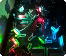
A multi-probe, multicolor, high-resolution miniaturized two-photon microscopy imaging platform — comprising multiple femtosecond lasers at different wavelengths and a series of miniature two-photon microscopes with different magnifications — meets the need for high-resolution multicolor brain imaging in freely behaving animals.
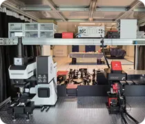
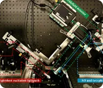
The system integrates three complementary imaging modalities, including single-photon light-sheet imaging, two-photon light-sheet imaging, and two-photon point-scanning microscopy.
Ultrasensitive Hessian structured illumination super-resolution microscope reduces phototoxicity in super-resolution imaging and enables the longest-duration super-resolution imaging. By exploiting the universal priors of spatiotemporal continuity and relative sparsity of fluorescence signals, a sparse deconvolution approach achieves unprecedented photon-to-resolution conversion efficiency. The system enables live-cell super-resolution imaging with 60 nm resolution, 564 Hz imaging speed, and an extended imaging duration exceeding 1 hour.
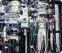
The 200 kV photoemission ultrafast cryo-transmission electron microscope is equipment developed by Prof. Li Jianqi’s research group at the Institute of Physics, Chinese Academy of Sciences. By integrating the high temporal resolution (picosecond-level) of the ultrafast laser system with the high spatial resolution (information resolution ≤0.14 nm) of the transmission electron microscope, it achieves in situ observation of complex transient structural dynamic processes in biological materials with high spatiotemporal resolution.
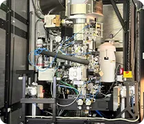
The 300 kV field-emission transmission electron microscope integrates a customized high-sensitivity X-ray energy-dispersive spectrometer, a high-speed energy-filtered direct electron detection system, a high-contrast pole piece, an integrated differential phase contrast (iDPC) detector, an EELS, a liquid specimen holder, and other components. It can perform multiple imaging modes and multimodal detection on biomacromolecule solution samples as well as cryo-sectioned cell and tissue samples.
The Biomedical Computing Platform of National Biomedical Imaging Center provides high-performance scientific computing and AI computing capabilities, along with massive data storage capacity and convenient access interfaces. It supports cutting-edge research and technological innovation in the field of biomedical imaging.
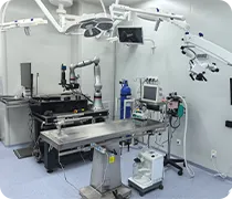
An auxiliary research platform supporting multimodal biomedical imaging experiments, equipment operation, and translational research workflows.